
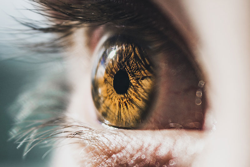
Special Eye Testing
When should children have their eyes examined?
No-Puff Glaucoma Testing
What’s the worst part of going to the eye doctor? For most people, it’s the part of the appointment when the Optometrist shoots a scary puff of air directly onto their eyes. Thankfully, you don’t need to put up with this inconvenient and uncomfortable test any longer!
Visual Field Testing
Visual Field testing can help save vision because it is another test used to diagnose or rule out glaucoma and other neurological disorders that affect vision. This simple, but effective service has saved lives by detecting various medical conditions such as strokes, brain tumors, and other neurological defects.
Eidon Fundus Photography, or Retinal Digital Imaging
A high-definition digital image of the retinal area helps your eye doctor diagnose and manage eye diseases in the delicate retinal area. Damage to the delicate structures of the retina is often the first sign of systemic diseases such as MS, diabetes and others. The retina is the “window to the body” and routine retinal imaging can help your eye doctor monitor the changes in your eye health from year to year.
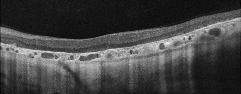
Optical Coherence Tomographer (OCT)
Our OCT helps us better manage glaucoma and diseases of the retina because this technology allows the eye doctor to see the deep tissue layers in the eye. Similar to ultrasound, this diagnostic technique employs light rather than sound waves to achieve higher resolution pictures of the structural layers of the back of the eye. These high-definition images are the only way that they can actually see beneath the surface to the nerve fiber layers where damage occurs. Up until now, eye doctors had to use other tests to indicate damage in this critical area of sight. Common eye diseases such macular degeneration, diabetic retinopathy, and glaucoma are detected early by the OCT when the diseases can be more effectively treated.